About this image
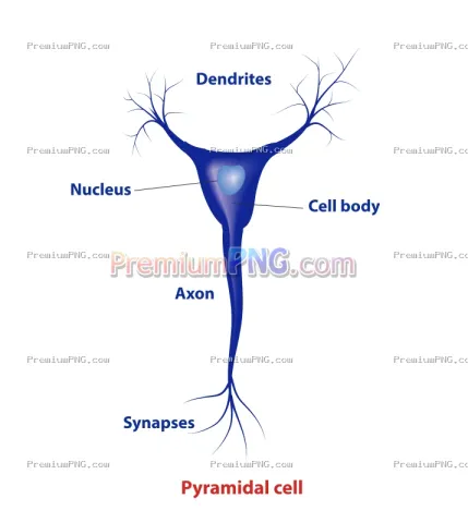
Pyramidal cell anatomy is a vector EPS illustration in the Science › biology collection on PremiumPNG.com. As a vector file it can be scaled to any size, from an icon to a large print, without losing sharpness. Contributed by KKT Madhusanka.
How you can use it
Open and edit the EPS file in any vector design program to change colours, shapes and size for logos, packaging, posters and print projects. It is licensed for personal and commercial projects under the Commercial License. Read the license details.
Similar Images
Visual & Style Match


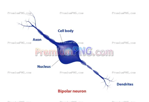
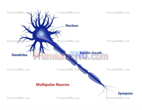

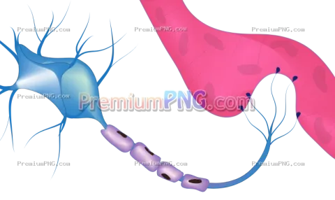


Pyramidal cell anatomy
High-quality Pyramidal Cell Anatomy vector EPS illustration featuring a detailed scientific diagram of a pyramidal neuron and its anatomical structures, including the pyramidal cell body (soma), nucleus, apical dendrite, basal dendrites, axon, axon hillock, and dendritic branches. This educational neuroscience illustration demonstrates the characteristic structure and morphology of pyramidal neurons commonly found in the cerebral cortex and hippocampus. Ideal for neuroanatomy studies, neuroscience research, biology textbooks, medical education, neurology presentations, classroom teaching, scientific publications, and academic learning materials. Scalable and editable vector EPS artwork suitable for professional printing, educational posters, digital presentations, and biomedical illustrations.
- Full-resolution EPS vector, no watermark
- Commercial License · lifetime
- No subscription, does not use plan limits
You must be logged in to download files.
Login to DownloadImage information
| Category | : Science |
| Subcategory | : biology |
| Image Type | : EPS |
| Resolution | : Scalable |
| File Size | : 6.26 MB |
| Uploaded Date | : October 28, 2026 |
| License | : Commercial License |
| Downloads | : 0 |
Contributor credit line